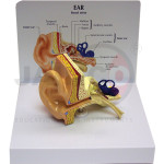
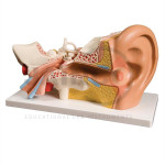
Anatomical Ear Model Manufacturer,Supplier and Exporter in India
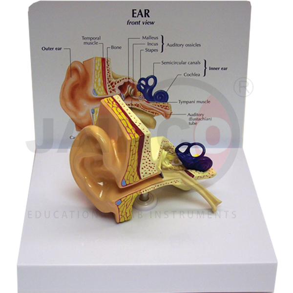
Product Code : JA-EM-324
This anatomical ear model is a one-piece, life-size cast of the human ear. It shows the outer, middle and inner ear together. The inner ear has the semicircular canals and the cochlea. The middle ear shows the auditory ossicles and the tympanic membrane with the middle-ear muscles. A card comes with the model.
Specifications of the life-size anatomical ear model
| Product code | JA-EM-324 |
|---|---|
| Size | Life size; 3.75 x 2.5 x 2.75 in |
| Construction | One piece |
| Regions shown | Outer, middle and inner ear |
| Inner ear | Semicircular canals; cochlea |
| Middle ear | Auditory ossicles; tympanic membrane; middle-ear muscles |
| Card | 6.5 x 5.25 in |
| Supplied by | JaincoLab |
Teaching the structure of the ear with the life-size model
- Tracing the path of sound. Students point in turn to the tympanic membrane, the ossicles and the cochlea. Hang an Ear, Nose, Throat Chart beside the model so they can match each part to its label.
- Simple hearing tests in physiology practicals. Strike a tuning fork and hold it beside the ear, then press its stem on the bone behind it. The model shows how sound reaches the cochlea through air and through bone.
- Placing the ear inside the head. The middle and inner ear sit within the temporal bone. Point this out on a Human Skull Model before passing the ear model round.
- Sense organ lessons in Class 10 and 11 biology. Set the ear model next to a Human Eye Model and compare how each organ turns a stimulus into nerve signals.
Frequently Asked Questions
Can the ossicles or the inner ear be taken out of this model?
No. The model is cast in one piece, so no parts lift out. The ossicles, tympanic membrane, cochlea and semicircular canals all stay in place on one surface. Choose a multi-part ear model if students need to take it apart.
How large is the model compared with a real ear?
It is life size, at 3.75 x 2.5 x 2.75 inches, so students see the real proportions of the ear. The ossicles are very small at this scale. Students see them most clearly when they look at the model up close in small groups.
In what order do the ossicles pass sound to the inner ear?
The malleus comes first, as it is joined to the tympanic membrane. It passes the vibration to the incus, and the incus passes it to the stapes. The stapes then pushes on the oval window of the inner ear.
Which inner ear part is for hearing and which is for balance?
The cochlea is for hearing. It is the coiled tube that turns vibration into nerve signals. The semicircular canals are for balance, as they sense turning movements of the head. The model shows both, so students can compare their shapes.
Where does the middle ear end and the inner ear begin?
The tympanic membrane closes off the outer ear. Behind it lies the air-filled middle ear with its three ossicles. The inner ear begins at the oval window, where the stapes sits, and holds the cochlea and the semicircular canals.
Other ear models in the ENT range
See the ENT Models range and more teaching models from JaincoLab, an anatomical models manufacturer in India.
For a quotation, send the product name and the quantity you need through our contact page. For tender or project supply, see our tenders page.